Cells massage their nucleus to repair DNA, revealing a possible cancer vulnerability
Cells soften and massage their nucleus to encourage the repair of broken DNA, research led by Barts Cancer Institute, Queen Mary University of London, has discovered.
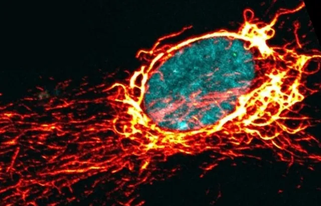
Microscope image of a cell nucleus and cytoskeleton, captured by the Godinho lab. DNA is shown in cyan, with components of the cytoskeleton in red and yellow.
Using cells grown in the laboratory, the researchers uncovered a previously unknown role for movement within the cell in DNA repair. Their findings could point towards new ways to make cancer cells more sensitive to existing treatments that damage DNA or disrupt its repair.
The study, published today, 28 October, in Nature Cell Biology, reveals how cells temporarily soften their nucleus following DNA damage. This allows fibres from the cell’s internal skeleton to push and pull on the nucleus, creating tiny fluctuations in its shape that wobble its contents.
The research was led by Professor Susana Godinho and co-first authors Dr Sam Wallis and Dr Wai Yiu Tse at Barts Cancer Institute, Queen Mary University of London.
“One way to picture it is that the cell massages its nucleus to help the DNA inside move around more freely, helping it to search for a matching sequence it can use to repair the break,” explains Professor Godinho, Senior author of the new study and Group Leader at Barts Cancer Institute, Queen Mary University of London. “When we disrupted this process, cells accumulated more DNA damage, stopped dividing, and became more sensitive to cancer drugs such as cisplatin and PARP inhibitors. That raises an interesting question: could blocking this pathway make cancer cells more sensitive to existing treatments?”
How broken DNA finds its match
Our DNA is damaged every day by factors ranging from ultraviolet light to metabolic processes taking place inside our own cells. If this damage is not repaired correctly, it can cause permanent changes to the genome and contribute to ageing, cancer and cell death.
Cells have therefore evolved sophisticated mechanisms for repairing their DNA. These include a process called homologous recombination, which fixes particularly serious breaks where both strands of the DNA double helix have been severed.
To repair this type of break accurately, the damaged DNA must locate an intact copy of the same sequence, usually on a recently copied piece of DNA. This presents the cell with a major challenge: finding one matching sequence among the billions of DNA letters packed inside the nucleus. Once the match is found, the intact DNA acts as a template, allowing the cell to replace the missing or damaged genetic information.
This repair process is particularly important in cancer. Many cancer cells divide rapidly and have unstable DNA, causing them to accumulate DNA damage. They can therefore become heavily reliant on DNA repair to survive – a dependency that some cancer treatments are designed to exploit. Understanding how cancer cells repair their DNA, and finding ways to disrupt it, may reveal new possibilities for treatment.
Uncaging the nucleus
The nucleus is normally surrounded by a rigid cage made from fibres of a protein called vimentin. This protective structure helps the nucleus retain its shape and withstand physical stress.
In the new study, the researchers discovered that DNA damage starts a chain reaction of events that temporarily disassembles this cage.
First, an enzyme called αTAT1 modifies microtubules – fibres that form part of the cell’s internal skeleton. The altered microtubules act like tracks along which sections of the vimentin cage are carried away from the nucleus.
With this protective brace loosened, the nucleus becomes softer and more flexible. Even small movements of the microtubules can then gently push and pull it out of shape, causing the nucleus to deform and its contents to wobble.
Crucially, the study showed why this movement matters for DNA repair: it helps broken DNA to move back and forth, allowing it to explore a larger area of the nucleus. This increases its chances of locating the matching sequence needed for repair.
Why this movement matters
When the researchers genetically removed αTAT1 from cells, it prevented the changes to microtubules that normally follow DNA damage. Without αTAT1, the nucleus remained stiffer and damaged DNA was less able to search for a matching sequence.
As a result, homologous recombination became less efficient, unrepaired breaks began to build up, and affected cells paused their cycles of cell division.
Removing αTAT1 also made cells more sensitive to three cancer drugs: cisplatin, which damages DNA, and the PARP inhibitors olaparib and talazoparib, which exploit weaknesses in cancer cells’ ability to repair DNA.
“What makes αTAT1 interesting is that cells can survive without it under normal conditions, but become much more dependent on it when they are under stress from DNA damage. Blocking it might therefore present an option to make some cancer cells more sensitive to existing treatments,” Professor Godinho explains.
However, the researchers emphasise that this possibility is still at an early stage of investigation. Currently, a suitable drug is not available that can inhibit αTAT1 for this purpose, and further research is needed to identify which cancers depend on the pathway and establish whether it could be targeted safely and effectively.
The team now plans to investigate how this pathway works in living organisms and will also explore whether certain types of double-stranded DNA breaks are more reliant on this process than others.
The findings challenge the often static picture of biology presented in textbooks. They show that DNA repair is a dynamic process shaped not only by molecules, but also by motion and physical forces at play within the cell.
Co-first author Dr Sam Wallis Postdoctoral Researcher and Microscopy Facility Manager at Barts Cancer Institute, says:
“This story really demonstrates how observing cell biology in real time can drive new discoveries. It shows that biological processes are not just molecular pathways but that cells also undergo mechanical and physical changes that play a fundamental role in how they function. Our hope is that this new understanding can one day be exploited therapeutically to enhance existing treatments and improve patient outcomes.”
The study was carried out in collaboration with researchers at University College London, the Francis Crick Institute, the Babraham Institute and the Princess Máxima Center for Pediatric Oncology and was supported by funding from Wellcome, Cancer Research UK and UKRI. See the original paper for a full list of funding sources.
To find out more about Queen Mary's research, sign up for Queen Mary University of London's newsletter.
Related items
 News story:
From AI to space medicine: William Harvey Day showcases research and innovation
27 October 2026